Research
welcome
Projects of the research group Morphology and Structural Analysis
We focus the investigations on life cycle and reproduction and culture and rearing of cnidarians. Furthermore, very important issues are examinations of equilibrium sense organs of rhopaliophoran medusae and examinations of stinging cells. These aspects are used for the elucidation of taxonomical and phylogenetical relationships of Cnidaria.
Equilibrium sense organs
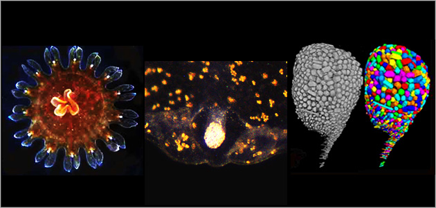
Ephyrae and medusae live predominantly pelagical in the free water column. They consist of 95-98 % water and their soft-body pass through changing phases of growth and degrowth during their development. Specimens have usually marginal sense organs at the bell margin, which provide an orientation in space for the specimen. The marginal organs of Scyphozoa and Cubozoa (rhopalia) are very complex and bear an statocyste at their distal end, which serves in cooperation with sensoric cilia as an equilibrium sense organ. The statocyst consists of cells, that contain anorganical crystals (statoliths). These crystals are the only hard-structur of the specimen and consist in all so far examined rhopaliophoran medusae of calciumsulfat subhydrate. Analyses of the statolith material (single crystal analysis) are performed in cooperation with Dr. Frank Hoffmann (department of chemistry / University of Hamburg). The statocyst develops differently in diverse systematic groups but is always increasing in size during development of the specimens. Statoliths are reacting weaker to changing invironmental influences like food availability than the soft-bodies and are therefore better suitable for growth and age analyses. An age analysis based on the jellyfish morphology only is not possible because of recurring shirinking phases , but desirable, because it could give informations about place of origin and could allow a forcast of mass occurances. The number of crystals within one statocyst is low at the beginning of development, increases during the development and can reach several thousands in a fully grown medusa. The shape of the statoliths can be very different and the shape of existing statoliths can change during development also. For example, in cubomedusae fuse the independant single statoliths into one large homogenous statolith. Statoliths and statocysts have also very specific charcteristics, that can be used for taxonimic purposes. In the research group are various methods for statolith examination established like lightmicroscopy (slide scanning), confocal laserscanning microscopy, scanning and transfocal microscopy, microtomography and 3D-reconstruction with AVIZO. Microtomography is performed in cooperation with Dr. Peter Michalik and Jakob Krieger (University of Greifswald). A very important basis for our investigations is the breeding and rearing of living specimens. We investigate particularly statocysts and statoliths of the Rhopaliophora to evalute the data collection for taxonomic and phylogenetical relationships. Also the development of an age determination is pursuded for the five large jellyfish species occuring in the North-Sea. The research on equilibrium sense organs is carried out in cooperation with Dr. Sabine Holst (DZMB).
Stinging cells
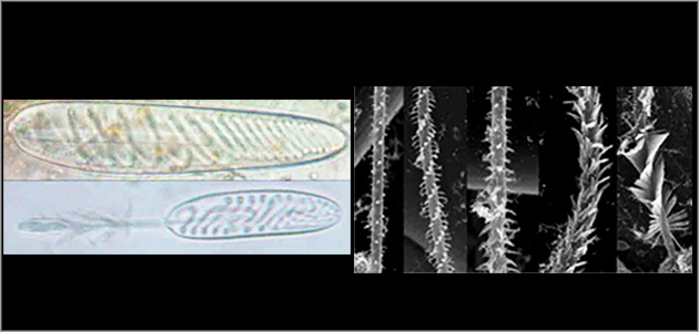
The stinging cells (cnidocytes) are existing in all cnidarien specimens. The capsules (cnidocysts), which are located inside the stinging cells exist in many different characteristic types that can be used for taxonomic differentiation. So far, nematocysts are examined mostly with lightmicroscopical methods, which allow a categorisation for capusle shape, shaft typ and tubule type. Investigation of more defined details, e.g. the spikes on the tubuuls are limited with the lightmicroscope. Therefore additional investigations using scanning electron micrsocopy are necessary. Also comprehensive and detailed investigations of all life cycle stages (planula, polyp, ephyra, medusa) are scarce, but indispensable for the description of the species specific cnidom. We investigate cnidocysts of Scyphozoa with lightmicroscopy and scanning elektron microscopy in collaboration with Dr. Sabine Holst (DZMB).
Life cycle and reproduction
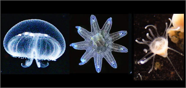
Alternations of generations is primarily present in three cnidarian taxa. Their polyp generation is mostly small, inconspicuous and sessile, and produces asexually the predomiantly pelagic medusa generation. The medusae produce sexually the planula larvae, which transform into polyps after settlement. Even though this life cycle is typical for most Hydrozoa, Scyphozoa and Cubozoa, there are some exceptions existing. In some species (e.g. Pelagia noctiluca, Periphylla periphylla) is the polyp generation lacking and planula larvae develope directly into medusae. Other species have a medusa generation, which is reduced to different degrees. This reduction has an influence on the reproduction mode; the specimens are hermaphrodites (Nausithoe eumedusoides) , reproduce parthenogenetically (Thecoscyphus zibrowii) or pure vegetatively (Nausithoe planulophora). Recently it became known that not only cubozoan medusae but also some scyphozoan medusae perform special mating behaviours (Periphylla periphylla). Life cycles and reproduction modes of many species are still not completely discovered and are an important part of interest in this research group. Basis for the investigations is the steady rearing of polyp cultures (approx. 30 species) and the breeding of epyrae and medusae. We exchange information and specimens with different institutions like the Multimar Wattforum, the Aquarium Berlin, the Zoo Duisburg und other researchers like Prof. André C. Morandini (Universität Sao Paulo), to optimize breeding protocols and to keep the species long-lasting in culture.
Culture and rearing
The rearing and care for living cultures is an important basis for education. We keep five different Aurelia species to a large extent for courses. Also wie breed other animal groups like protozoans e.g.Amoeba proteus, Blepharisma japonicum, Paramecium caudatum, Thecamoeba, Chlorococcum sp. and Nanochloropsis salina as well as metazans like polychaets
(e.g. Ophryotrocha sp.), oligochaets, plathelminths, rotatorians (Brachionus plicatilis), gastropods (Helix pomatia, Biomphalaria glabrata), crustaceans (Tisbe holothuriae, Artemia salina, Daphnia magna u.a.) and waterbears (Hypsibius durjardini).
